It’s magical enough to stand inside the Pacific Seas Aquarium and gaze at shimmering scales, cruising sea turtles or impossibly long wolf eels.
But with x-ray vision, a whole new underwater world reveals itself.
Thanks to our veterinary team, who regularly take radiographs of our Pacific Seas Aquarium animals to check on their health and care for them, we can all see what lies just their skin or scales.
And it’s truly amazing.
Azul, green sea turtle
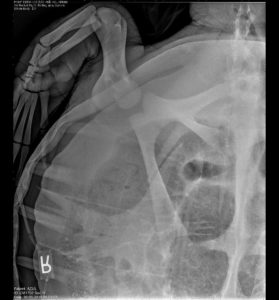
This x-ray clearly sees through Azul’s shell to his scapula (the shoulder blade, in a human), and on up to his humerus (upper arm bone) and on to the digits of his front right flipper.
“With this x-ray we are able to look at his skeletal structure, his bones and joints, as part of his overall health screening,” explains Dr. Karen Wolf, lead veterinarian. “Azul’s radiograph is normal, and will serve as a baseline for any changes in the future.”
Sunny, green sea turtle (close-up)
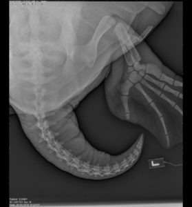
This close-up clearly shows Sunny’s left rear leg, including the large femur, the smaller tibiotarsus and five digits of his flipper, alongside a curvy tail.
“What I love about this is how you can really compare it to a human foot,” says Wolf.
Turtles use their flippers to manipulate their environment, like humans use hands and feet: as well as swimming and “walking” while on sand, they can push food closer and leverage or hold other objects (like coral) to better access prey.
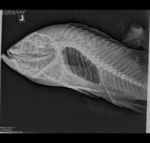
Buddy, California sheephead
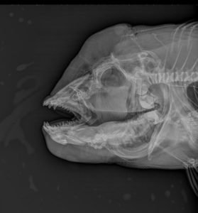
Buddy lives in the Coastal Kelp Forest habitat and is a firm favorite with Zoo guests and aquarists alike.
Looking past his distinctive pink-and-gray outside, though, you can clearly see the sharp canine teeth and big jaw he uses to demolish prey. You can also see that the bulbous shape of his head is due to fleshy tissue that makes up his forehead, rather than the skull itself.
Scalloped hammerhead shark
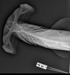
This x-ray highlights the beautiful, elongated, almost surreal head structure (cephalofoil) of a hammerhead shark.
“Eye sockets at either end give excellent binocular vision,” says Wolf. “You can also see the gills, slender spine and delicate fins.”
Black rockfish
The Northwest Waters exhibit teems with rockfish of all colors and patterns – but one thing they all have in common is a swim bladder, a gas-filled organ that helps the fish control its buoyancy in the water.
In this x-ray the swim bladder is clearly visible as a large dark space under the spine.
Flounder
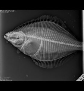
“I love this radiograph, because it shows this fish’s unique anatomy,” says Wolf.
Evolved to live on the sea floor, flounders are flat and asymmetrical. Here you can clearly see the pectoral fin and two eye sockets on the top side.
As a hatchling, flounders have one eye on each side, but as they mature one eye migrates around. You can also see the fish’s numerous delicate spines.
Wolf eel and moray eel
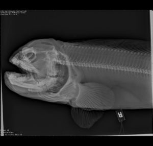
Our wolf eel can be spotted lurking in a rock den in the Under the Narrows habitat, while moray eels need warmer water like our Coastal Kelp Forest.
Although the wolf eel is technically a fish (it has pectoral fins), both have long spines, small heads and sharp teeth.
But morays have something special – an extra set of jaws.
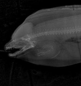
“The pharyngeal jaws are a second set deeper inside that help the eel hold onto prey,” Wolf explains.
In this x-ray, look for a fainter, ghostly jaw-shape just to the right of the main jaws that gives the fish an alien appearance.
(NOTE: The L and R appearing in these x-rays are tags laid next to the animal while radiographing to enable accurate identification of left and right side.)