Black cats, pumpkins, ghosts, skulls, and skeletons symbolize the Halloween season. We asked Point Defiance Zoo & Aquarium’s animal care team, head veterinarian Dr. Karen Wolf, and the veterinary technicians to give us an “inside” look at a few of the Zoo’s animals and their not-so-spooky skeletons. The veterinarians regularly take radiographs or X-rays of the animals to check their health and care for them.
Walnut the Beaver
In this X-ray, you can see Walnut the beaver’s long incisor teeth. A beaver’s incisors grow continuously throughout its life and are worn down through daily use. These teeth are also self-sharpening.
Scooter the Armadillo
In this radiograph, you can see Scooter the armadillo’s armored skin. In the wild, this armor protects armadillos from predators.

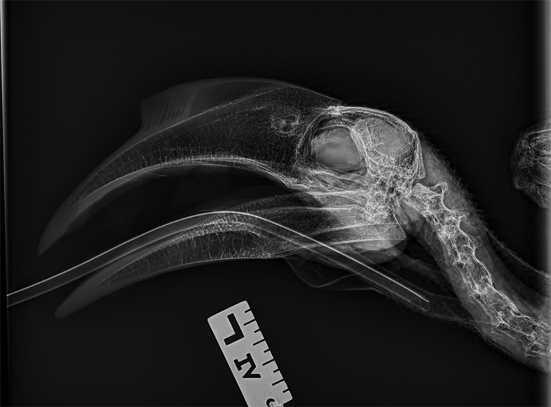
Pilai the Hornbill
In this picture, you can see Pilai the hornbill showing off her impressively large beak. Hornbills use their bills to snatch prey.
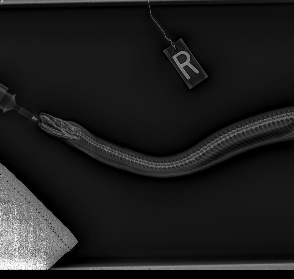
Scarlet the Rainbow Boa
In this picture, you can see all of the delicate vertebrae and rib characteristics of snakes. You can also see the boa’s many sharp teeth angled backward, helping the snake keep its prey headed in the direction of its stomach.
Yuna the Malayan Tapir
This radiograph displays the long snout of Malayan tapir Yuna, which is ideal for foraging. It also reveals her specialized shearing teeth adapted for chewing vines, leaves, twigs, and fruit.

Titan the Rhinoceros Iguana
Rhinoceros iguanas are named for the horn-like protrusions on their heads. Interestingly, these unique lumps don’t appear in radiographs because they’re not composed of bone and are actually large scales. 
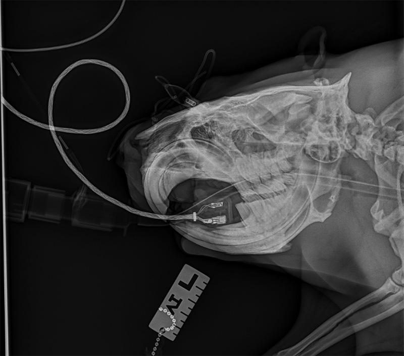
Orchid the Clouded Leopard
This radiograph provides an incredible image of Orchid, the clouded leopard. Clouded leopards have some of the largest canine teeth of any wild cat, proportional to their body size.

Rockfish
While there are many types of rockfish, they typically all have spines on their heads, bulging eyes, and a large mouth. In this picture, you can see the giant eye socket and individual spines.
Xena the African Pouched Rat
This X-ray shows our African pouched rat’s strong jaw and large cheek pouches. These pouches can hold nearly as much food as the rat’s body weight—perfect for storing snacks to enjoy later.

Splendid the Magnificent Tree Frog
Here you can see the long leg bones of the magnificent tree frog. Their limbs and flexible toes are designed for gripping branches and making impressive leaps through their rainforest home.

Naledi the Meerkat
In this X-ray, you can spot the slender skeleton of a meerkat, built for digging and quick movement. Their upright posture helps them watch for predators while their family forages below.

Waffle the Damaraland Mole Rat
This radiograph highlights the Damaraland mole rat’s long front teeth and streamlined body, which are perfect adaptations for underground life. These social rodents work together to dig intricate tunnels beneath the surface.

Kanak the New Caledonia Giant Gecko
This image shows the New Caledonia giant gecko’s lightweight bones and long tail that help it balance as it climbs. These nocturnal lizards are skilled climbers that rely on their sticky toe pads for grip.

Piranha
This X-ray reveals the piranha’s sharp, triangular teeth and powerful jaw. In the wild, these fish use their specialized teeth to slice food efficiently, playing an essential role in their river ecosystems.

Larry the Puffin
This radiograph highlights the puffin’s specialized wing bones, which function like flippers when diving underwater. Thanks to this dual-purpose design, puffins are agile fliers in the air and efficient swimmers below the surface.

Wolf Eel
The wolf eel’s elongated skeleton is designed for slipping into rocky crevices along the seafloor. Its strong jaws help it crush hard-shelled prey like crabs and sea urchins, making it an expert reef hunter.

Tilli the Aardvark
This radiograph focuses on Tilli the aardvark’s skull, showing her elongated snout and specialized teeth. Unlike most mammals, aardvarks don’t have front incisors or canines. Instead, peg-like teeth in the back of their jaws continuously grow and wear down as they dig for ants and termites.

Laerke the Polar Bear
In this X-ray, you can see the powerful skull of polar bear Laerke. Her large canines and strong jawbones are designed for gripping and tearing through thick prey like seals. The shape of her skull also helps support strong neck muscles needed for hunting in Arctic waters.

Baby Bean the Lowland Anoa
This radiograph captures the skull of our Lowland anoa, Baby Bean, including her sturdy jaw and horn cores. As one of the smallest wild cattle species, her skull shows how well-built anoa are for foraging on tough vegetation and maneuvering through dense forests.

Boo the Bamboo Shark
This radiograph shows the bamboo shark’s gills and jaw structure. Its cartilage-based skeleton keeps it light and flexible, and its gills allow it to pump water over itself and breathe even while resting on the sea floor.
